Slovak scientists join forces in the fight against staphylococcal infection
Bacteria are among the smallest yet most dangerous adversaries in medicine. While some are harmless, others can cause serious infections where early diagnosis is crucial for successful treatment. A team of Slovak scientists from the Slovak Academy of Sciences is therefore exploring how to detect the presence of bacteria directly in tissue—quickly, accurately, and without the need for invasive procedures. Their research combines confocal Raman microscopy, photodynamic therapy, and data analysis using a supercomputer.
Challenge: Recognizing whether tissue is infected with bacteria is not always straightforward. In the early stages of infection, the differences between healthy and damaged cells often cannot be detected even under a microscope. Although traditional biochemical tests can confirm the presence of bacteria, they are usually time-consuming and require sample collection.
Solution: To identify subtle differences between healthy and infected tissue, the researchers decided to combine experimental measurements with advanced data processing. Raman spectra obtained from different depths and regions of the tissue contained an enormous amount of information that could not be reliably evaluated using conventional visual methods.
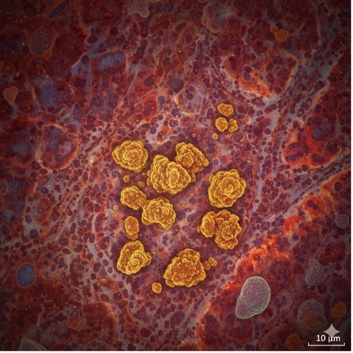
The scientists therefore sought to verify whether this method could reliably distinguish healthy tissue from tissue infected with Staphylococcus aureus one of the most common causes of skin and mucous membrane inflammations. At the same time, the researchers focused on monitoring the effectiveness of photodynamic therapy—an experimental treatment based on carbon quantum dots that, when exposed to blue visible light, destroy bacteria without harming healthy cells.
Use of HPC Infrastructure
The team employed a mathematical analysis based on the Euclidean cosine of the squares of the first differentiated values, which enables the comparison of similarities between spectra after their transformation. This method eliminates background interference, highlights chemical changes in the tissue structure, and allows precise identification of differences caused by the presence of bacteria or the effects of treatment.
The computational power of a supercomputer was used to process the extensive datasets. Thanks to parallel data processing, it was possible to rapidly analyze hundreds of measurements from different tissue layers and visualize their similarities in a clear results matrix. Such an approach would be virtually impossible through manual evaluation.
The solution was developed through close collaboration among experts from multiple disciplines—biology, physics, materials research, and computational science. Reconstructed skin tissues were provided by the SK-NETVAL laboratories at the Institute of Experimental Pharmacology and Toxicology of the Centre of Experimental Medicine, Slovak Academy of Sciences (SAS), which also performed the exposure to the tested substances. The photodynamic treatment was applied by the team from the Polymer Institute of SAS, and the Raman data were collected at the Institute of Physics of SAS in cooperation with the Centre for Advanced Material Application.
Results
The analysis of the spectral data revealed significant chemical differences between healthy and infected tissue that can be detected using Raman microscopy. Samples infected with Staphylococcus aureus showed distinct spectral characteristics at all analyzed depths.
The results were particularly interesting in the samples that underwent photodynamic treatment. After the application of carbon quantum dots and subsequent activation with blue light, the chemical spectra closely approached those of healthy tissue. This suggests that the treatment effectively suppresses bacterial infection without damaging the cells themselves.
The applied algorithm proved to be a reliable and fast tool for comparing spectral data. Thanks to its implementation in the HPC environment, it was possible to automatically process large volumes of measurements and evaluate the results objectively, without any subjective interference from the researcher.
Impact and Future Potential
The project has brought new insights into the potential use of light and data analysis in medical diagnostics. It has demonstrated that combining Raman microscopy with computational methods enables not only the identification of bacterial infection in tissue but also the monitoring of treatment effectiveness in real time.
In the future, this approach could be applied in the development of new antibacterial therapies or in preclinical drug testing, where it is essential to quickly and accurately assess structural changes in tissue without invasive procedures. The research team also plans to extend the methodology to other types of bacteria and tissues and to leverage the computing power of the supercomputer for testing advanced artificial intelligence algorithms that could further automate the analysis.
The project is proof that the integration of biomedicine, physics, materials research, and computational science opens new possibilities for disease diagnosis and treatment.
Slovak research teams are thus not only demonstrating their scientific excellence but also contributing to pushing the boundaries of modern medicine.

 Nový recept na skrotenie slnečnej energie 11 Jun - Umelá inteligencia v spojení s čistou fyzikou dokáže predpovedať silu slnečného žiarenia rýchlejšie a lacnejšie než kedykoľvek predtým